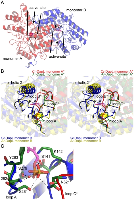
Figure 7. Location and orientation of the active-site of Cr-DapL.
A) Location of the two active-sites in the dimer, as highlighted by the position of a PLP molecule, taken from an overlay of Cr-DapL with Ar-DapL+PLP structure (2Z20),shown in stick form (magenta). PLP was not found in the active site of Cr-DapL. B) Stereoview of the active-site showing the loops that contribute residues to the active-site. Again PLP is added to the structure from an overlay with Ar-DapL+PLP structure (2Z20). The image overlays the monomers of Cr-DapL (blue and red) with that of the apo-Ar-DapL (yellow and green). In B) and C), the asterisk emphasizes loops that are contributed from the opposing monomer in the dimer. C) Bonding of residues in loops A and C* with the sulfate, which sits in the same position as the phosphate of PLP.

