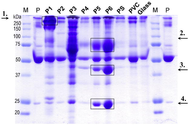
Figure 6.
Patterns of plasma proteins adsorbed onto 2 mg of polymer particles after a 30-min incubation in hirudin-plasma. The gel shows, from the far left: molecular marker (M), hirudin plasma (P) diluted 1/50; P1 through P6 are the synthesized polymers, followed by the reference polymers PS, PVC, and glass. To identify the proteins that were enriched on polymers P1, P5, and P6, Western blot analysis was used, and the results showed that P1 had a high affinity for C1q (≈410 kDa, arrow 1), whereas P5 and P6 were enriched in vitronectin (≈75 kDa, arrow 2), apolipoprotein AIV (≈45 kDa, arrow 3), and apolipoprotein AI (≈25 kDa, arrow 4) on their surfaces. In contrast, HSA (migrating at ≈55–60 kDa) was seen in similar amounts to all surfaces. Comparable results were obtained when the particles were incubated in EDTA-plasma or serum (data not shown).

