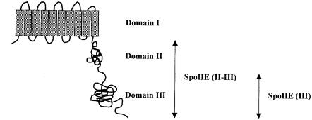
Fig. 1. Topological model for the multi-domain structure of SpoIIE protein. The membrane-spanning segments are shown as grey rectangles and the soluble part of the protein is shown as two adjacent globules. The cytoplasmic domain of SpoIIE consists of domains II and III (residues 326–827) and is referred to as SpoIIE (II–III) in the text. Domain III (residues 568–827) contains the PP2C phosphatase and is referred to as SpoIIE (III).

An official website of the United States government
Here's how you know
Official websites use .gov
A
.gov website belongs to an official
government organization in the United States.
Secure .gov websites use HTTPS
A lock (
) or https:// means you've safely
connected to the .gov website. Share sensitive
information only on official, secure websites.
