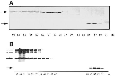
Fig. 3. Interaction of SpoIIE, SpoIIE (II–III) and SpoIIE (III). (A) SDS–PAGE of SpoIIE (II–III) and SpoIIE (III) after separation on a Superdex 200 gel filtration column and staining with Coomassie Blue. SpoIIE (II–III) was eluted between 59 and 77 ml, corresponding to an apparent Mr between 300 and 50 kDa. SpoIIE (III) was eluted between 84 and 90 ml, corresponding to an apparent Mr of ∼28 kDa. The upper and lower arrows indicate the position of SpoIIE (II–III) and SpoIIE (III), respectively. (B) Purified full-length SpoIIE (3.2 μM) and SpoIIE (III) (3 μM) were mixed and applied to a Superdex 200 gel filtration column equilibrated with buffer C. Elution was performed in the same buffer and fractions of 1 ml were collected and monitored by Western blotting with anti-SpoIIE antibodies. Full-length SpoIIE was eluted between 46 and 66 ml, corresponding to an apparent Mr between 700 and 170 kDa. SpoIIE (III) was eluted between 84 and 91 ml, corresponding to an apparent Mr of ∼28 kDa. The positions of full-length SpoIIE and SpoIIE (III) are indicated by solid arrows. The additional immunoreactive bands (indicated by dotted lines) correspond to dimeric and higher molecular mass (possibly tetrameric) forms of SpoIIE.

An official website of the United States government
Here's how you know
Official websites use .gov
A
.gov website belongs to an official
government organization in the United States.
Secure .gov websites use HTTPS
A lock (
) or https:// means you've safely
connected to the .gov website. Share sensitive
information only on official, secure websites.
