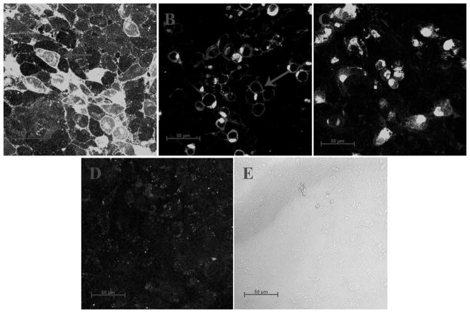
Figure 2.
Confocal microscopy images testing for cellular localization of truncated SNAT2-AcGFP fusion constructs. (A) SNAT2WT, (B) SNAT2Del C-ter, and (C) SNAT2Del TM11. Panel (D) shows control, non-transfected cells, and (E) is the bright-field control image, showing that cells were present. The arrows indicate regions of intense cell boundary fluorescence. AcGFP was attached in frame to the N-terminus of SNAT2.

