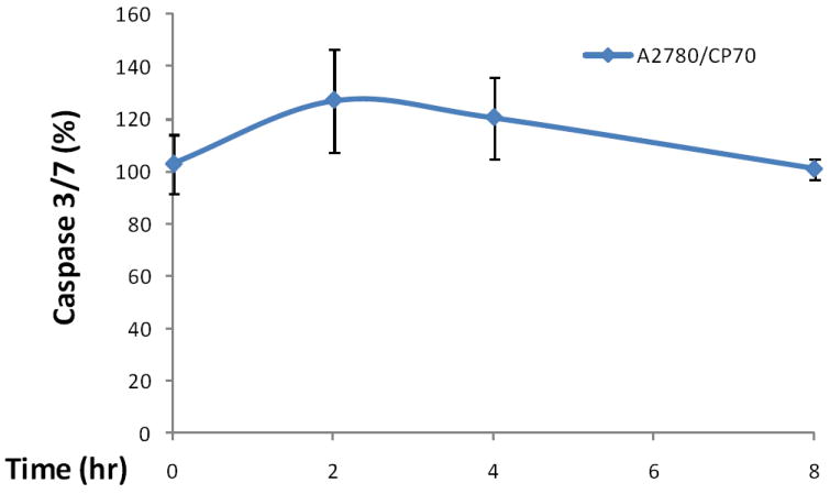
Figure 3. Time course of kaempferol-induced apoptosis in A2780/CP70 ovarian cancer cells.

A2780/CP70 ovarian cancer cells were seeded in 96-well plates at 10,000 cells/well and incubated overnight. The cells were treated with 0- or 80-μM kaempferol in triplicates for 0, 2, 4, or 8 hours. No-cell wells were included for background correction. At each time point, a plate was removed of culture medium and frozen in -80 °C until final analysis. Cells were thawed, lyzed with Passive Lysis Bufer, and analyzed for caspase 3/7 activities with a Caspase-Glo 3/7 Assay and total protein levels with a BCA assay. Caspase 3/7 activities were normalized by total protein levels, and the levels of kaempferol-treated cells were expressed as percentages of the controls for statistics. Data represent Means ± SE from 2 independent experiments.
