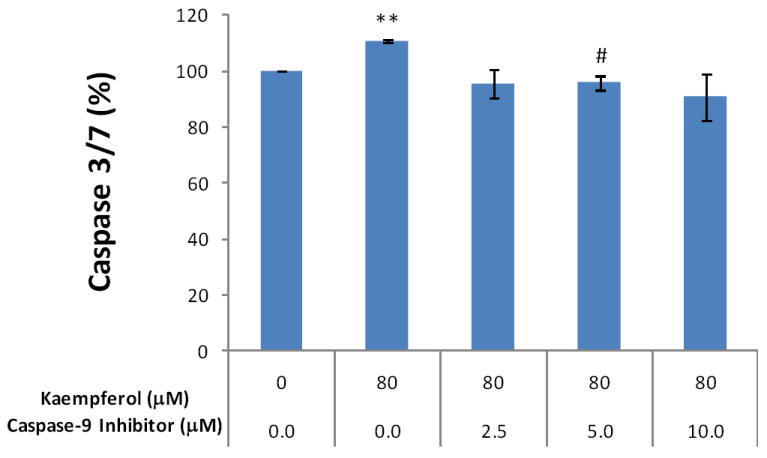
Figure 6. Kaempferol induces apoptosis through intrinsic pathway in OVCAR-3 ovarian cancer cells.

OVCAR-3 cells were seeded at 10,000 cells/well in 96-well plates and incubated overnight. The cells were pre-treated with various concentrations of a caspase-9 inhibitor (Z-LEHDFMK) or negative control (Z-FA-FMK) in triplicate for 24 hours, and treated with 0 or 80 μM of kaempferol for 2 hours. For the caspase 3/7 assay, 100 μl freshly prepared reagent was added to each well, incubated for 1 hour at RT, and 60 μl of the reaction mixture was transferred to glass tubes to measure luminescence. For cell number assay, 100 μl of freshly prepared AqueousOne reagent was added, incubated for 1 hour at RT, and measured at OD 560 with a microplate reader. Non-cell wells were included to measure background values for both assays. Caspase 3/7 values were adjusted by cell number values. Data represent Means ± SE from 2 independent experiments. **p<0.01 as compared to control. #p<0.05 as compared to the kaempferol treated control.
