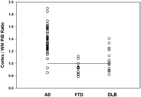
Figure 3.
Volume of interest PiB binding ratio of frontal cerebral cortex to subcortical white matter (WM). DVR ratios are depicted for each subject according to their qualitative image analysis-based classification. The horizontal line depicts the theoretically employed threshold of abnormality of 1.0. Based on the volume of interest threshold, there are two cases qualitatively classified as abnormal (Alzheimer’s disease) that fall below the quantitative threshold and two cases qualitatively classified as normal (frontotemporal dementia) that fall in the quantitatively abnormal range (see text for detailed discussion). AD = Alzheimer’s disease; DLB = dementia with Lewy bodies; FTD = frontotemporal dementia; WM = subcortical cerebral hemispheric white matter.

