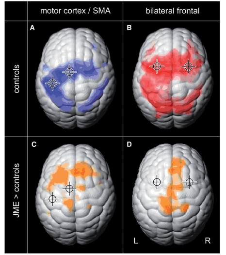
Figure 3.
Functional connectivity is increased in JME. Functional connectivity of the motor system and of the cognitive working memory system were analysed by two seed regions each (crosshair shows location of the seed region). Group connectivity maps of healthy controls show functionally connected cortical areas for the left motor cortex and SMA in blue (A, FDR, P < 10−6), and for the bilateral frontal working memory network in red (B, FDR, P < 10−6). Group difference maps in orange show increased functional connectivity in JME compared with controls: motor cortex and SMA are increasingly connected to prefrontal cortex (C, FDR, P < 0.001), and the bilateral working memory network shows increased connectivity to the medial central region, SMA and medial prefrontal areas (D, FDR, P < 0.005). There were no areas of increased connectivity in healthy controls.

