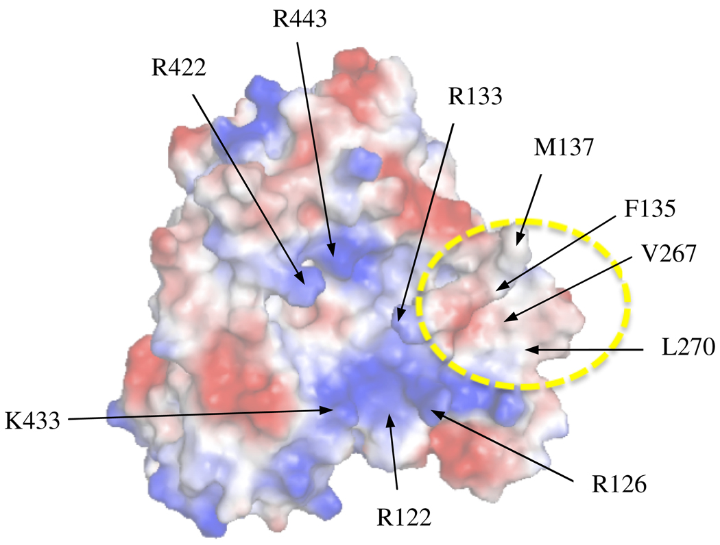
Figure 1.
Surface electrostatic plot of the proximal region of CYP2B4. Electrostatic surface of CYP2B4 as determined by APBS which shows the central basic residues proposed to be involved in CPR binding and a hydrophobic patch that includes V267 and L270 indicated by the dashed yellow circle. Red represents acidic residues while blue represents basic residues on the surface of CYP2B4.

