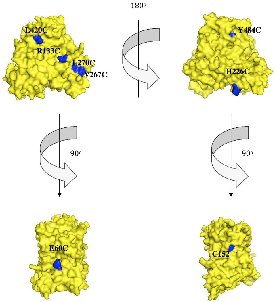
Figure 2.
Selection of solvent-exposed residues for FM labeling. van der Waal surface representations of CYP2B4 (PDB 1SUO) in the upper left and right corners display the proximal and distal regions of CYP2B4 respectively. The lower left and right structures represent regions perpendicular to the axis of the heme-thiolate ligation. The residues that were selected for cysteine mutagenesis and subsequent FM labeling are shown in blue, while the rest of the structure is shown in maize. Since WT CYP2B4 possesses two surface-exposed cysteines (C79 and C152) almost all our studies required generating a C79S/C152S template to which cysteine residues were introduced at sites of interest. However, residue C152 (lower right) was already in a position of interest and studying this site simply required replacing C79 with serine.

