Figure 2.
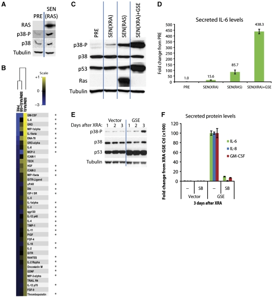
p38MAPK drives the amplified SASPs induced by RAS or p53 inactivation. (A) p38MAPK is phosphorylated during RASV12-induced senescence. PRE cells were infected with lentivirus expressing oncogenic RASV12, selected, and allowed to senesce (SEN(RAS)) for 10 days. Whole cell lysates were then collected and analysed by western blot. Presenescent controls (PRE) were infected with insertless vector. (B) p38MAPK inhibition suppresses the SEN(RAS) SASP. Cells were infected as described in (A). CM from PRE and SEN(RAS) cells were analysed by antibody arrays. +SB: p38MAPK was inhibited by SB203580 for 48 h before CM collection. Shown are the top 40 factors for which the SEN(RAS) level was significantly increased over PRE. PRE and SEN(RAS) values were averaged to generate the baseline. Heat map and dendrogram were generated as in Figure 1E. *: Factors significantly decreased by p38MAPK inhibition. (C) Amplified p38MAPK phosphorylation in SEN(RAS) cells and SEN(XRA) cells lacking functional p53 (SEN(XRA)+GSE). Cells were infected with lentivirus expressing GSE22 (GSE) or an insertless vector, selected, then irradiated (XRA) or infected with lentivirus expressing oncogenic RASV12 (RAS) and allowed to senesce. Whole cell lysates were analysed by western blot. (D) SEN(RAS) and SEN(XRA) cells lacking functional p53 secrete amplified IL-6 levels. Cells were treated as in (C), then CM were collected and analysed by ELISA. (E) p53 inactivation accelerates p38MAPK phosphorylation after XRA. Cells were infected with lentivirus lacking insert (Vector) or expressing GSE22 (GSE), selected, and irradiated. Whole cell lysates were collected at specified time points and analysed by western blot. (F) GSE-amplified levels of IL-6, IL-8, and GM-CSF are p38MAPK dependent. Cells were infected as in (E) and irradiated. CM were collected 3 days later and analysed by ELISA. SB: p38MAPK was inhibited by SB203580 for 48 h before CM collection.

