Fig. 2.
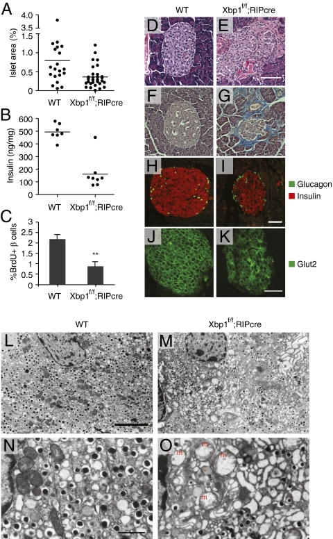
Histopathological analysis of islets of β-cell-specific Xbp1 KO mice. (A) Islet area relative to total pancreas. One random pancreas section per mouse was examined. Each dot represents an individual mouse of the indicated genotype of 4–6 mo of age. (B) Pancreatic insulin contents of 12- to 16-wk-old male mice. (C) BrdU-positive β-cells were counted after BrdU/insulin double staining. n = 4–5 mice per group. **P < 0.01. Pancreatic sections from WT and Xbp1f/f;RIP-cre mice were stained with (D and E) hematoxylin and eosin, (F and G) trichrome blue, (H and I) insulin and glucagon, and (J and K) Glut2 antibodies followed by fluorescence-conjugated secondary antibodies. (Scale bars, 100 μm.) (L–O) Islet sections were examined by TEM. [Scale bar, 5 μm (L and M); 1 μm (N and O).] The asterisk indicates an electron lucent granule. m, swollen mitochondria.

