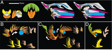
Fig. 5.
Comparison with extant lamprey and other conodont taxa. (A) (Left) Supraoral tooth (green) and lingual laminae (orange: transverse lamina; yellow: longitudinal laminae) of the lamprey G. australis. (Right) Sagittal sections of the lamprey head in protracted (middle) and retracted (right) positions. Red: muscles; cyan: cartilages. Redrawn after Hilliard et al. (23). (B–D) Proposed relative positions and movements of the elements of Ellisonia (B), Hibbardella (C), and Paracordylodus (D). Isolated S1–4 in lower rows. Color coding as in Fig. 2C; light orange: basal body. Modified, respectively, after Koike et al. (24), Nicoll (25), and Tolmacheva and Purnell (26). (B) M is missing.

