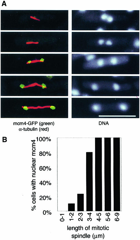
Fig. 4. mcm4 binds to chromatin during anaphase B. Cells from an asynchronous culture (P560) in YE were processed using the in situ chromatin binding procedure, after which cells were stained with anti-α-tubulin antibody. (A) The left images indicate cells in different stages of anaphase B, showing mitotic spindles (red) and chromatin-bound mcm4 (green). Bar = 10 μm. The right images show the corresponding DNA staining (DAPI). (B) Proportion of mitotic cells with mcm4-positive nuclei shown according to mitotic spindle length. On average, 12 cells were scored for each length class (range 5–20 cells).

An official website of the United States government
Here's how you know
Official websites use .gov
A
.gov website belongs to an official
government organization in the United States.
Secure .gov websites use HTTPS
A lock (
) or https:// means you've safely
connected to the .gov website. Share sensitive
information only on official, secure websites.
