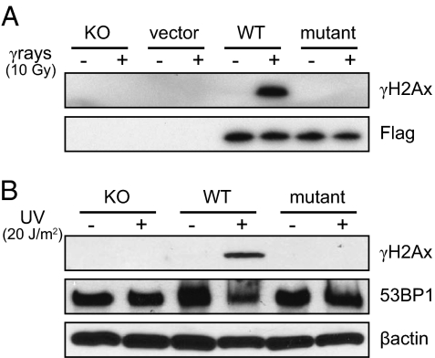
Fig. 1.
Expression of γH2Ax formation is restricted to cells transfected with wild-type H2Ax. (A) γ-Rays (10 Gy, 30 min); (B) UV (20 J⋅m−2, 4 h) cells. The Flag epitope indicates the presence of the H2Ax vector in both wild-type and mutant cell lines. 53BP1 is expressed in each cell type as this is a DSB binding protein that is constitutive but relocates to DSBs, and is used as an independent marker.

