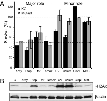
Fig. 4.
Relative sensitivity of H2Ax mouse knockout (black histograms) or mutant (gray histograms) following exposure to γ-rays, etoposide, rotenone, temozolamide, UV, cisplatin, and mitomycin C. (A) The doses required to kill 50% of the population (LD50) were recorded from multiple repeats of the survival curves in Figs. 2 and 4 [γ-rays n = 1 (Fig. 2A); Etop n-3; Rot n = 3; Temoz n = 6; UV n = 6; UV plus caffeine n = 6; Cispt n = 3; MitC n = 3] and means and SEs calculated. Lower dashed line indicates the relative sensitivity when the damage is predominantly double strand breaks, as for γ-rays and etoposide. The upper dashed line at 100% indicates the absence of any sensitization from inactivation of γH2Ax. (B) Western analysis of the levels of γH2Ax induced in cells with wild-type H2Ax 4 h after the start of exposure to an LD50 dose of each designated agent or 30 min in the case of γ-rays. Abbreviations: Etop, etoposide; Rot, rotenone; Temoz, temozolamide; Cispl,cisplatin; Mit C, mitomycin C.

