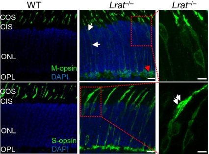
Fig. 1.
Mistrafficking and accumulation of cone opsins in P18 Lrat−/− retina. Lrat−/− and WT retinas were stained with M-opsin and S-opsin antibodies (green). Nuclei were stained with DAPI (blue). In Lrat−/− retina, mistrafficking of M-opsin is indicated by white arrows (Top and Middle). Mistrafficking of S-opsin is apparent. Right: Magnified views of two boxed regions from middle. Accumulation and aggregation of S-opsin in the inner segment of an Lrat−/− cone is indicated by white arrows. Small amount of aggregated M-opsin is indicated by the red arrow. No immunoreactivity was detected in various mouse retinal sections when control rabbit IgG was used for staining (SI Appendix, Fig. S12). COS, cone outer segment; CIS, cone inner segment; ONL, outer nuclear layer. (Scale bars: Right, 5 μm; 10 μm elsewhere.)

