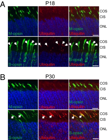
Fig. 3.
S-opsin and ubiquitin in Lrat−/− cones. (A) P18 and (B) P30 Lrat−/− retinal sections were double-labeled with antibodies against M-opsin or S-opsin (green) and ubiquitin (red). Aggregated S-opsin in P18 Lrat−/− cones colocalized with ubiquitin in A (white arrows). In P30 Lrat−/− retina, extensive colocalization between S-opsin and ubiquitin can be seen (white arrows) in B. Nuclei were stained with DAPI (blue). Labeling with isotype control mouse IgG1 (for ubiquitin antibody) or rabbit IgG (for M/S opsin antibodies) did not show nonspecific signals (SI Appendix, Fig. S12). (Scale bar: 10 μm.)

