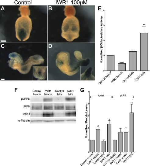
Fig. 4.
IWR-1 decreases or increases TOPGAL activity in wild-type embryos, depending on stage and tissue. (A–D) TOPGAL activity in cultured control (A and C) and IWR-1–treated (B and D) wild-type embryos that carry one copy of the TOPGAL transgene. (A and B) Posterior views of late headfold embryos after 18 h of culture, showing decreased expression of TOPGAL in the primitive streak of IWR-1–treated embryos. (C and D) Lateral views of 6-somite embryos after 18-h culture. At this stage, IWR-1–treated embryos showed reduced TOPGAL expression in the head, but increased expression in the primitive streak and PSM. The Insets in C and D are dorsal and ventral views of the primitive streak, respectively. The unusual shape of the tail is characteristic of embryos treated with the tankyrase inhibitors. (Scale bars, 100 μm.) (E) β-Galactosidase activity in anterior (head) and posterior (tail) halves of control and IWR-1–treated embryos, normalized to the activity in control heads. β-Galactosidase activity was measured in three independent experiments, with six to eight embryos per condition in each experiment. (F) Representative Western blots showing the increased level of Axin1 in embryos treated with 100 μM IWR-1 compared with DMSO controls. Embryos treated with IWR-1 also have a higher level of pLRP6 in the tails. (G) Analysis of Western blot data from six independent experiments in which four to eight embryos were cultured per condition in each experiment. Protein levels were normalized to α-tubulin, and values were normalized to protein levels in DMSO-treated heads. Axin1 levels were increased 1.90-fold in IWR-1–treated heads (P < 0.05) and 2.33-fold in treated tails (P < 0.05). The change in pLRP6 levels in treated heads was variable and not significant, whereas the 3.26-fold increase in IWR-1–treated tails was significant (P < 0.01).

