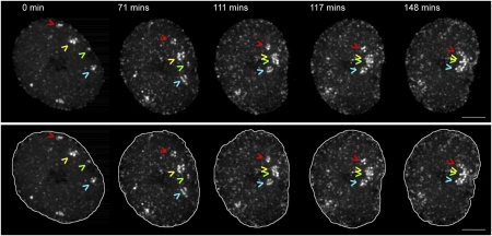
Fig. 1.
RCs move and coalesce inside the infected cell nucleus. A cell infected with ICP8-GFP virus was imaged in 4D starting at 4 hpi. The time series is displayed as a maximum intensity projection. Arrowheads point to four RCs that coalesce during the course of the movie. Lower panel shows the same time series with nuclear outlines. (Scale bar: 5 μm.)

