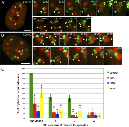
Fig. 4.
RCs coalesce at nuclear speckles. (A and B) Cells were transfected with a plasmid encoding mCh-SC35 for 24 h, and then, they were infected with ICP8-GFP virus and imaged at 4 hpi. Four major types of RC movement (arrowheads) relative to speckles (asterisks) are shown in rows 1–3. Rows 1a and 1b are high-magnification time-series images of the cell shown in A, and they show RCs (green) that move alone either to a speckle (red; 1a) or another RC (1b). Rows 2 and 3 are high-magnification time-series images of the cell in B, and they represent RCs that move on speckles or with speckles, respectively. Time stamps in A and B show hour:minute:second postinfection. Time stamps in rows 1–3 show minute:second (at 4 hpi) (Movie S2). (Scale bar: 5 μm.) (C) mCh-SC35 transfected cells were treated with control medium, lat A, or BDM at 3 hpi or with α-amanitin at 4 hpi, and they were imaged at 4 hpi. Numbers of RC coalescence events and types 1–3 movement were determined manually. The data are presented as mean percentage ± SEM per nucleus (∼7 nuclei and ∼44 RCs were analyzed per treatment). Asterisks mark values that are statistically different from control medium-treated cells (P < 0.05) (Movies S2, S3, S4, and S5).

