Fig. 1.
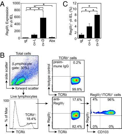
Intestinal microbiota direct RegIIIγ expression in small-intestinal γδ IEL. (A) RegIIIγ mRNA was quantified by Q-PCR of isolated small-intestinal γδ IEL from germfree (gf), conventionally raised (cv-L), conventionalized (cv-D), or antibiotic-treated (Abx) mice (n = 5 mice per group; representative of two experiments). (B) γδ IEL express RegIIIγ protein. Flow cytometry was performed on total small-intestinal IEL populations from cv-L mice. Intracellular staining was carried out with anti-RegIIIγ antibodies (9), and the gating strategy is shown. TCRδ+/RegIIIγ+ cells were analyzed to verify expression of the lymphocyte marker CD103. (C) Percentages of RegIIIγ+ γδ IEL were determined in gf, cv-L, and cv-D mice using the strategy shown in B. n = 5 mice per group; data are from two independent experiments. Error bars represent ±SEM. *P = 0.05.

