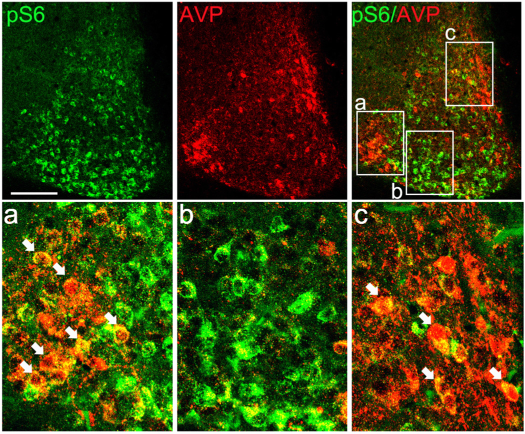
Figure 2. Dual immunolabeling for pS6 and arginine vasopressin (AVP) in the SCN.
Animals were dark-adapted for 2 d and killed at CT12. Upper row: representative confocal immunofluorescent images of pS6 (green, left panel) and AVP (red, middle panel). The merged image (right panel) reveals largely non-overlapping expression patterns. Framed regions a, b and c are presented as magnified panels in the lower row. Arrows indicate cellular-level colocalization (denoted by a yellow hue) of the two antigens. Scale bars: 100 microns.

