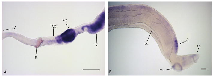
Figure 2.
argonaute localization in adult female (A) and male (B) Schistosoma mansoni worms. ago2 was developed for 30 min in males and 3 hours in females. argonaute is localized to the gonads (testes, ovary) and vitellaria of S. mansoni, with the highest expression in the posterior ovary (PO). Abbreviations: A, anterior end of worm; AO, anterior ovary; E, egg; GC, gynecophoral canal; OS, oral sucker; PO, posterior ovary; T, testes; V, vitellaria; VS, ventral sucker. Images obtained with a Zeiss Axio Star plus microscope. Scale Bars: 100 μm.

