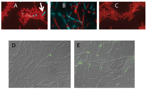
Fig. 2.
Heterogeneous expression of eps-cfp and spoIIE-gfp in mecA and pKD93 strains. Strains were grown in LB and sampled at T1. Panels A, B and C present typical fields from the wild-type (BD4621), mecA::erm (BD4642) and pKD93 (BD4643) strains respectively. Cell bodies were stained with propidium iodide and pseudocolored red. CFP fluorescence was pseudocolored cyan and overlayed on the propidium iodide channel. For panels D and E, a strain expressing spoIIE-gfp was grown in DSM to T-1. Images showing GFP fluorescence were overlayed on DIC images. Panels D and E show images from strains with the wild-type (PP480) and mecA::erm (PP479) backgrounds respectively.

