
Figure 5.
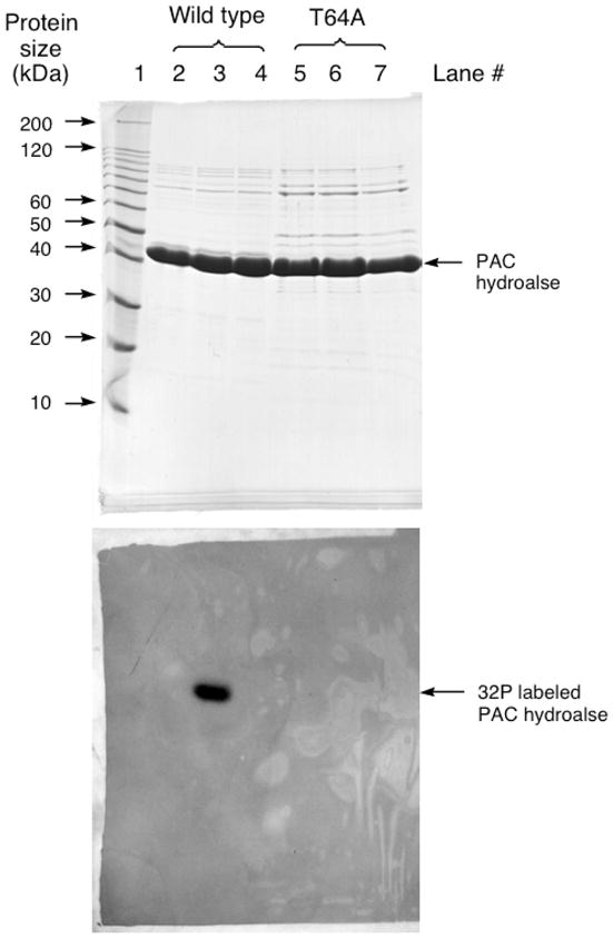
SDS-PAGE analysis of PAc hydrolase autophosphorylation with [γ 32P]ATP. The top panel is the Coomassie blue stained gel and the bottom panel is the audioradiograph of the gel. Lane 1: Protein molecular weight standards. Lane 2: The reaction of [γ 32P]ATP with acid-denatured wild-type PAc hydrolase. Lane 3: The reaction of [γ 32P]ATP with wild-type PAc hydrolase. Lane 4: The reaction of [γ 32P]ATP with wild-type PAc hydrolase in the presence of 2 mM phosphonoformate. Lane 5: The reaction of [γ 32P]ATP with acid-denatured T64A PAc hydrolase. Lane 6: The reaction of [γ 32P]ATP with T64A PAc hydrolase. Lane 7: The reaction of [γ 32P]ATP with T64A PAc hydrolase in the presence of 2 mM phosphonoformate.