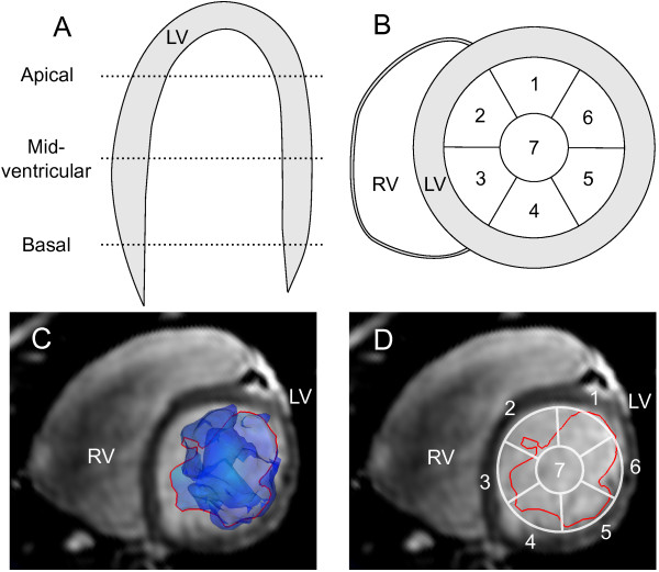
Figure 3.
Division of the LV lumen into segments. In panel A the LV lumen is divided into apical, mid-ventricular and basal parts along the long-axis. Panel B shows how each short-axis part is divided into seven segments, where six (labels 1-6) are located along the endocardial border adjusted from the AHA standard [29], and the seventh (label 7) in the center of the lumen. Panel C shows a representative slice and Volume Tracking surface (blue transparent surface). The intersection of the Volume Tracking surface with the short-axis slice is shown as a red line. Panel D shows the intersection in red and the segment model from panel B in white. LV = left ventricle, RV = right ventricle, 1-7 = segment numbers.

