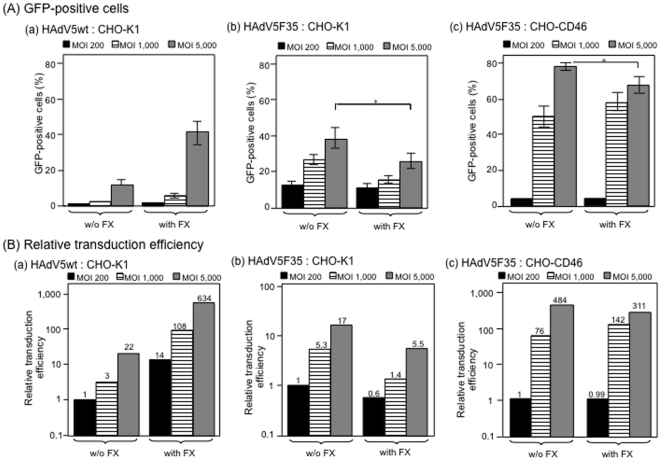
Figure 5. Comparison of transduction efficiency of (a, b) CHO-K1 cells or (c) CHO-CD46 by (a) HAdV5wt, and (b, c) chimeric HAdV5F35 vectors at different MOI (200, 1,000 or 5,000 vp/cell) in the absence (w/o) or presence of (with) FX (8 µg/ml).
Results were expressed as (A) the percentage of GFP-positive cells, or (B) relative transduction efficiency (RTE; refer to the legend to Fig. 2). In B, the number on top of each bar corresponded to the fold increase in RTE, with the 1-value attributed to the TE of CHO-K1 or CHO-CD46 cells transduced by HAdV5F35 at MOI 200. Note that RTE of CHO-K1 cells with HAdV5F35 was lower in the presence of FX than in the absence of FX, at all MOI tested.

