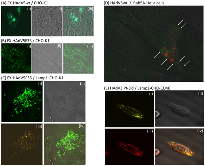
Figure 7. Confocal microscopy of live cells transduced by adenoviral vector particles or capsid components (penton dodecamers).
(A–C), Confocal microscopy of live cells (CHO-K1) transduced by Alexa-488-labeled adenoviral vectors, used at 10,000 vp/cell and complexed with FX (8 µg/ml). (A) HAdV5wt, 30 min pi; (B, C) HAdV5F35, 3 h pi. (i), Green channel image; (ii), phase contrast; (iii), merge of (i) and (ii). In (C), CHO-K1 cells were transduced by recombinant baculoviral vector expressing RFP-tagged, late endosome marker Lamp1 protein, 24 h before incubation with HAdV5F35 vector. (i), Green channel image; (ii), phase contrast; (iii) orange channel; (iv), merge of (i) and (iii). (D) Live HeLa cells transduced by recombinant baculovirus expressing RFP-tagged, early endosome marker Rab5A protein, were incubated 24 h later with Alexa-488-labeled HAdV5wt particles without FX, at 10,000 vp/cell and 37°C. Picture shown was taken at 20 min after incubation with HAdV5wt. Note that most of the virus signal is weak and diffuse, but some green fluorescent dots are visible within the cytoplasm (white arrows). (E), Live CHO-CD46 cells transduced by recombinant baculovirus expressing RFP-tagged, late endosome marker Lamp1 protein, were incubated 24 h later with Cy5-labeled HAdV3 penton dodecahedrons (Pt-Dd) at 37°C. Picture shown was taken at 60 min after incubation with Pt-Dd. (i), Cy5 channel; (ii), phase contrast image; (iii) RFP channel; (iv), merge of (i) and (iii).

