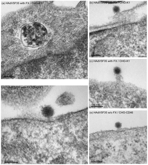
Figure 9. Electron microscopy of CHO-K1 cells (a–d) incubated with HAdV5F35 at 10,000 vp/cell in the presence of FX (8 µg/ml), and harvested after 2 h at 37°C.
(a), Representative CHO-K1 cell section showing a cytoplasmic vesicle containing abundant electron dense material. (b–d), Cell surface-bound HAdV5F35 particles. (e), CHO-CD46 cells incubated with HAdV5F35 in the absence of FX (w/o FX). Note the difference in size and sharpness of the viral contour between HAdV5F35 particles seen in (e) and in (b–d).

