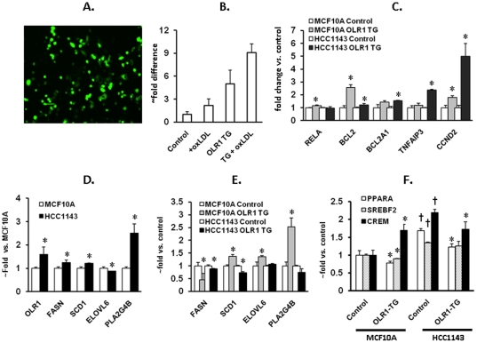
Figure 3. Effects of OLR1 overexpression on transcription of genes involved in apoptosis, proliferation and lipogenesis in MCF10a and HCC1143 cells.
These cells were transfected with either empty vector or OLR1 cDNA (Origene, Rockville, MD) using Lipofectamine 2000 (Invitrogen). Transfection efficiency (70–80%) was evaluated using GFP vector. RNA was extracted 48 hours post-transfection, converted into cDNA and the expression of genes was determined by quantitative PCR. A. Efficiency of transfection (cells transfected with GFP vector). B. Quantitative PCR plot. Note the enhancement of OLR1 expression in both control and OLR1-transfected cultures in response to ox-LDL. C. Expression of genes involved in apoptosis and proliferation. In order to stimulate OLR1 associated signaling requiring OLR1-ligand interaction, OLR1 transfected cells were treated with 40 µg/ml ox-LDL for 24 hours; graphs represent comparison with untreated control cells transfected with empty vector; D. Basal expression of OLR1, PLA2G4B and lipogenesis genes in normal human mammary epithelial cells (MCF10A) and breast cancer cells (HCC1143); E. Expression of OLR1, PLA2G4B and lipogenesis genes in MCF10a and HCC1143 cells transfected with OLR1 treated according to the protocol described above. F. Expression of lipogenesis transcription factors in MCF10a and HCC1143 cells transfected with OLR1 and treated according to the protocol described above. All experiments were conducted in triplicates. (*) p<0.05 compared to respective control; (†) – p<0.05 compared to MCF10A.

