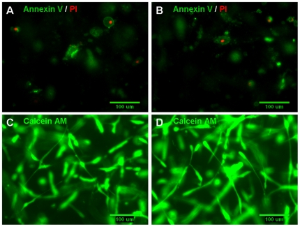
Figure 5. Exposure to shearing forces did not induce cell apoptosis or necrosis.
At the end of the migration period, both non-sheared cells (in control gels; A, C) and cells in gels exposed to 0.55 dynes/cm2 shear stress (B, D) were stained either by the Vybrant Apoptosis Assay Kit no. 2 or by Calcein AM. (A, B) There was no evidence of apoptosis being induced in the U87 cells as a result of exposure to the higher levels of shearing forces in this experiment; apoptotic cells were stained with Alexa Fluor 488 annexin V (green) and necrotic cells were stained with propidium iodide (red). (C, D) Calcein AM (green) staining indicates that a majority of the cells remained viable and cell morphology was normal in both gels containing non-sheared U87 cells and cells exposed to 0.55 dynes/cm2 shear stress for four hours.

