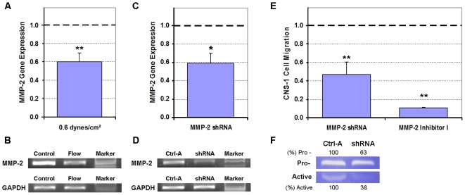
Figure 8. Shear-induced suppression of CNS-1 cell invasive potential was dependent on MMP-2 as determined by PCR, shRNA gene knockdown, and MMP-2 Inhibitor I.
All numerical data were normalized to their respective controls (1.0). (A) RT-qPCR data indicated that MMP-2 gene expression was downregulated in response to CNS-1 cells being exposed to 0.55 dynes/cm2 shear stress. (B) Gel electrophoresis of representative samples confirmed that the MMP-2 gene was suppressed in CNS-1 cell suspensions in response to flow (sheared cases) when compared to cells in control (non-sheared) gels. (C, D) RT-qPCR and gel electrophoresis of representative samples confirmed gene silencing of MMP-2 in CNS-1 cells transfected with the MMP-2 shRNA compared to cells transfected with the control (Ctrl-A) vector. (E) Migration of CNS-1 cells was suppressed when either MMP-2 gene expression was knocked down or when activated MMP-2 was targeted by MMP-2 Inhibitor I. (F) Gelatin zymography of conditioned media collected from the wells of inserts containing CNS-1 cells transfected with the MMP-2 shRNA confirmed that both pro- and active MMP-2 were down regulated compared to cells transfected with the control vector. Quantifications for MMP-2 expression are presented as percentage of their respective controls. Data presented as mean±SEM. Note: * p<0.05; ** p<0.005.

