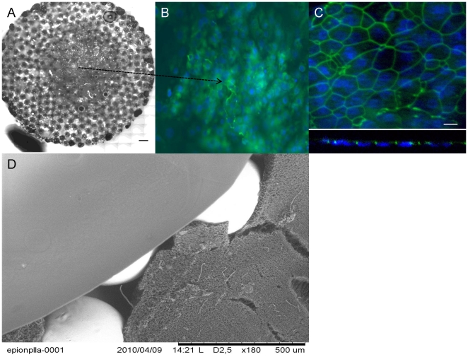
Figure 5. Overall structure of the hybrid implant and epithelialization.
a) Collation of a full surface of a circular PLLA/Ti hybrid implant, seeded with freshly isolated Human respiratory epithelial cells isolated from nasal polyps(Scale bar: 500 µm). c) DAPI and anti-ZO1 stainings showed monolayer formation on the hybrid implant surface (Day 19). c) Confocal images on x–y plane and Z-section confirms the development of strong cell-cell contacts. d) SEM image of the overall hybrid structure, showing the titanium beads, the micropous body and the top film layer.

