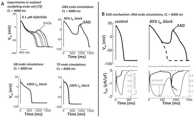
Figure 11. Early afterdepolarizations (EADs).
A) Top left) Experiments in isolated nonfailing human endo myocytes from Guo et al.[73] showed EADs with slow pacing (CL = 4000 ms) in the presence of IKr block (0.1 µM dofetilide, ∼85% IKr block[74], reproduce with permission). Top right) Following the experimental protocol of Guo et al. (CL = 4000 ms, 85% IKr block) the ORd model accurately showed a single large EAD. Bottom) GB (left) and TP (right) models failed to generate EADs (CL = 4000 ms, even with 100% IKr block). B) EAD mechanism. APs are on top. ICaL (black) and ICaL recovery gate (gray) are below. Slow pacing alone (CL = 4000 ms) did not cause an EAD (left). Slow pacing plus IKr block (85%) caused an EAD (solid lines, right). The EAD was depolarized by ICaL reactivation during the slowly repolarizing AP plateau (solid lines, solid arrows). When ICaL recovery was prevented, the EAD was eliminated (dashed lines and dashed arrow).

