Figure 4.
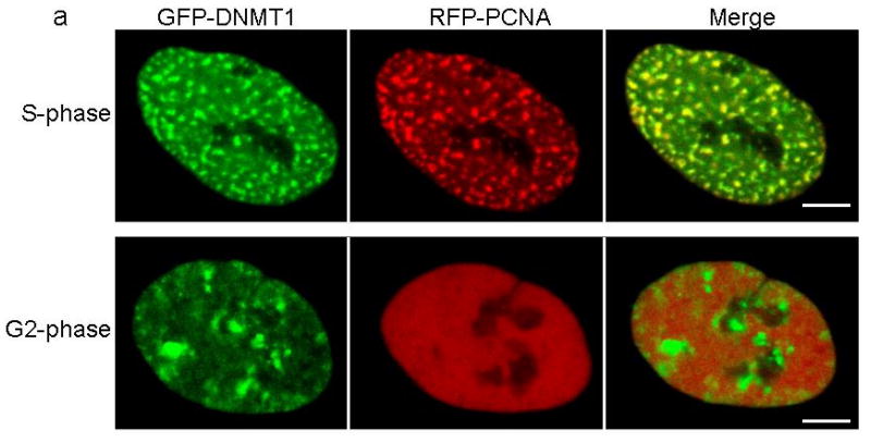
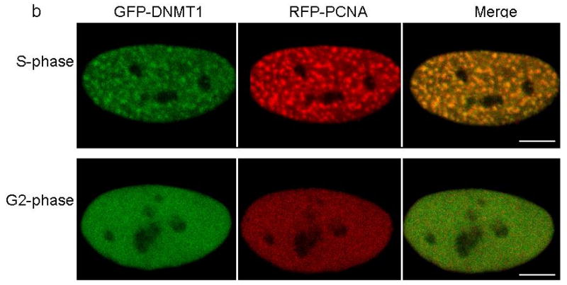
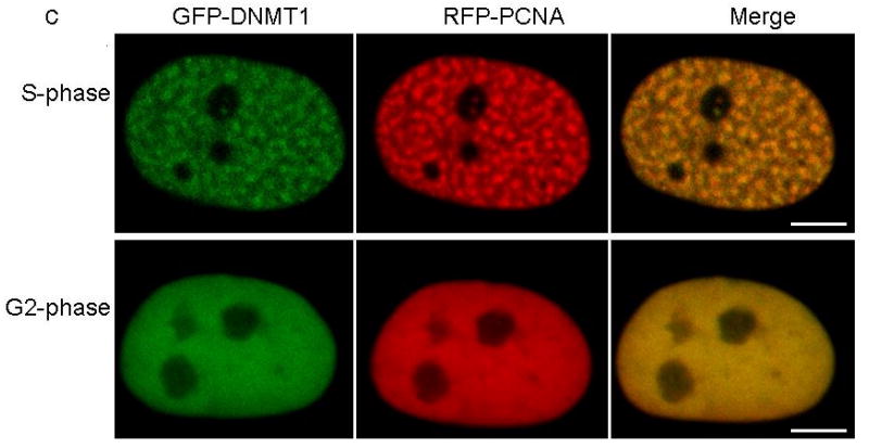
Confocal microscopy was performed using HeLa cells co-transfected with plasmids containing RFP-PCNA and full length (a) GFP-wild type DNMT1, (b) GFP-p.Tyr495Cys-DNMT1 or (c) GFP-p.Asp490Glu-Pro491Tyr-DNMT1. Wild type and mutant DNMT1 appear green in the right panels, PCNA appears red in the middle panels and merged images are shown in the left panels. Scale bar, 5um. Cell cycles are deciphered from the pattern of RFP-PCNA. In S phase, PCNA is present at the toroidal structures of the replication foci; in G2 phase, PCNA shows diffused pattern in the nucleus. In panel a, wild type DNMT1 co-localizes with PCNA at replication foci during both S phase and binds to heterochromatin during G2 phase. In panel b and c, p.Tyr495Cys-DNMT1 and p.Asp490GLu-Pro491Tyr-DNMT1 localize along with PCNA at the replication foci during S phase but did not show binding of heterochromatin during G2 phase.


