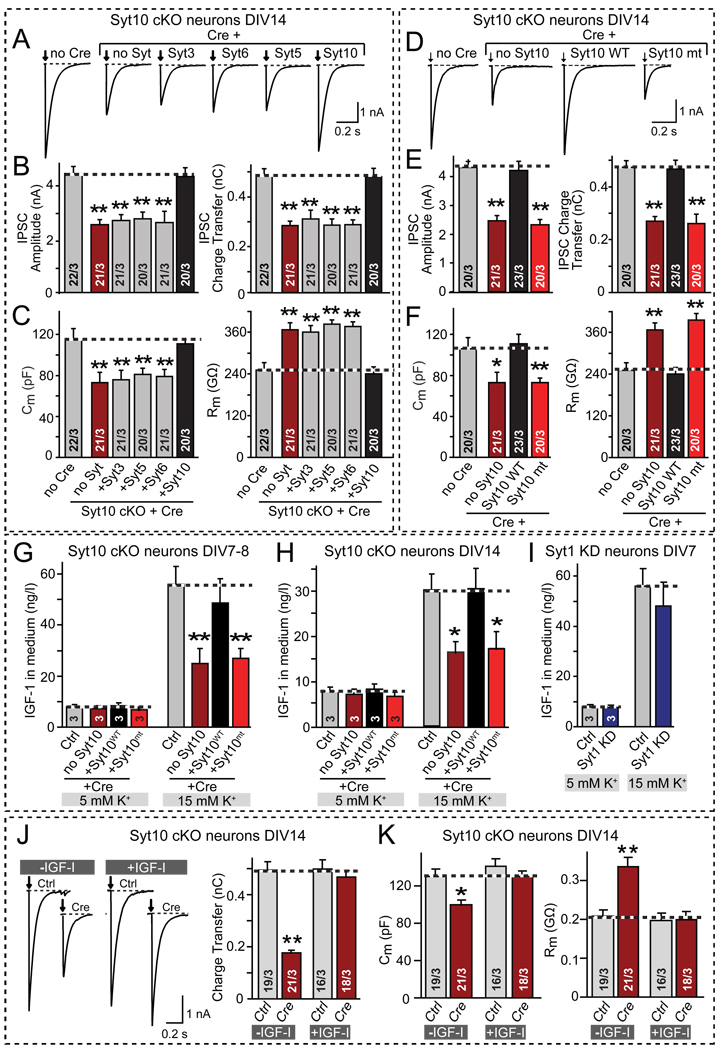
Figure 5. Syt10 functions as a specific Ca2+-sensor for IGF-1 secretion in cultured olfactory bulb neurons.
A–C. Only Syt10 but not the closely related synaptotagmin isoforms (Syt3, Syt5, and Syt6) rescue the Syt10 KO phenotype. Olfactory bulb neurons cultured from conditional Syt10 KO mice (Syt10 cKO) were infected with control (Ctrl) or Cre-recombinase expressing lentiviruses (Cre) that express the indicated synaptotagmins. Panels depict representative traces of evoked IPSCs (A) and summary graphs of the amplitude (B, left) and charge transfer of evoked IPSCs (B, right), and of the capacitance (C, left) and input resistance (C, right) monitored in mitral neurons (see also Fig. S3A).
D–F. Same as A–C, except that neurons were rescued with wild-type (Syt10WT) or mutant Syt10 in which the Ca2+-binding sites of both C2-domains were abolished (Syt10mt). For a description of the mutation, see Fig. S3B.
G & H. Measurements of depolarization-induced IGF-1 secretion by cultured olfactory bulb neurons after stimulation with 5 or 15 mM K+ for 1 hr at DIV7–8 (G) or DIV14 (H). Neurons cultured from conditional Syt10 KO mice were infected with lentiviruses expressing inactive (Ctrl) or active cre-recombinase (Cre), the latter without (no Syt10) or with co-expression of wild-type (Syt10WT) or Ca2+-binding site mutant Syt10 (Syt10mt). The IGF-1 concentration in the medium was measured by ELISA. Note that the secretory capacity decreases with the age of the culture, but the effect of the Syt10 KO remains the same. (see Fig. S3C for ELISA standardization).
I. Same as G and H, except that wild-type olfactory bulb neurons without or with shRNA-dependent knockdown of Syt1 (Syt1 KD) were analyzed.
J. Representative traces (left; arrows = action potentials) and summary graphs of the charge transfer (right) of IPSCs monitored in mitral neurons from conditional Syt10 KO mice that were infected with control (Ctrl) or cre-recombinase-expressing lentivirus (Cre) at DIV2, and maintained in the absence or presence of 50 µg/l synthetic IGF-1 (~6.5 nM) added to the medium from DIV7 onwards (see Fig. S3E for IPSC amplitudes).
K. Summary graphs of the the neuronal capacitance (left) and input resistance (right) of mitral neurons treated as described in J.
All summary graphs depict means ± SEMs; number of cells and number of independent cultures analyzed is shown in individual bars except for G–I, in which the number of independent culture experiments is indicated in the bars. Statistical analyses were performed by Student's t-test comparing the cre-recombinase treated neurons to control neurons (*=p<0.05; **=p<0.01).

