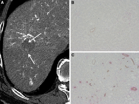
Fig. 2.
Early HCC (high-grade DN with a well-differentiated HCC focus). A CT during hepatic arteriography (CTHA) shows entirely hypodense nodule (arrow b) with a slightly hyperdense focus (arrow c). B Double immunohistochemical staining for CD34 and αSMA of DN portion (arrow b in A) shows no definite expression of sinusoidal capillarization and unpaired arteries. C Double immunohistochemical staining of HCC portion corresponding (arrow c in A) shows expression of sinusoidal capillarization and unpaired arteries.

