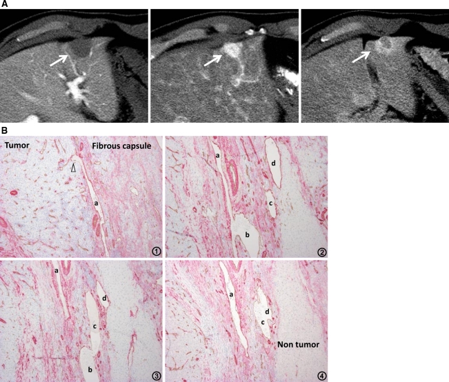
Fig. 7.
Moderately differentiated HCC with pseudocapsule formation. A CTAP shows an entirely hypodense nodule (left, arrow). CTHA demonstrates it as an entirely hyperdense nodule on early phase (middle, arrow) with thick corona enhancement on late phase (right, arrow). B Serial specimens from 1 to 4 with double immunohistochemical staining for CD34 and αSMA show the communication between intra-capsular portal venules (a–d) and intratumoral blood sinusoids (arrowheads) (×100).

