
Figure 5.
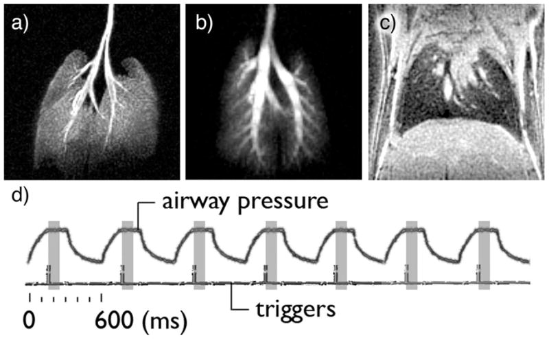
a) maximum intensity projection (MIP) of a volume image set acquired on a BALB/c mouse ventilated with oxygen and hyperpolarized 3He. b) MIP from a different Balb/C mouse, ventilated with oxygen and hyperpolarized 129Xe. c) 313-micrometer thick 1H slice through the lungs. The sharp edge at the base of the lungs suggests that the ventilator accurately delivered the same tidal volume —and repositionned the diaphragm at the same location— 4000 consecutive times, the number of breathing cycles required to acquire the image. d) The recorded airway pressure increases during the 150-ms inhalation, followed by a 150 ms-long plateau, indicating breath hold. The airway pressure decreases during the 300 ms expiration. Each breathing cycle is identical and lasts 600 ms. The imaging triggers, located 160-ms into the breathing cycle, indicate the start of the partial acquisition of the image, which lasts 100ms (shaded rectangle).