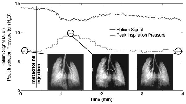
Figure 7.
a) During a 4-minute-long challenge induced by 250 μg/kg dose of the broncho-constrictor methacholine in a mouse, a series of 20 two-dimensional images were acquired consecutively, one image every 12 second. The magnitude of the 3He signal (k=0) remains largely constant (full line), although an exponential decay is apparent due to relaxation within the Tedlar bag. The dashed line shows the peak inspiration pressure, revealing a clear increase in airway resistance after the injection of methacholine, 24 s after the start of the experiment. An image of the mouse lungs is shown before methacholine injection (left), during broncho-constriction (middle) where the larger airways have decreased in diameter (arrows), and after recovery (right).

