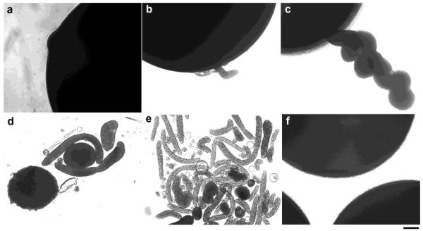
Figure 1. Tubulo-vesicular structures extruded from oocytes overexpressing wild type (WT) MLN1.
Formation of these structures at 2-4 days after injection of WT-MLN1 cRNA (a and b), and at later stages 5-8 days after injection (c-e), are shown. No such structures were observed in control oocytes (injected with H2O) (f) incubated for the same periods of time (1-8 days after injection). Scale bar: 100 μm.

