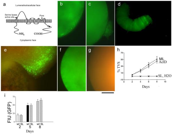
Figure 2. Formation of TVS is deficient in oocytes expressing MLN1 with a mutated serine lipase active site (SL-MLN1).
This site is located on the lumenal/extracellular side of MLN1 near its putative 1st transmembrane domain, while the pore region is between the putative 5th and 6th transmembrane domains (a). The oocyte shown in (b) has been injected with GFP-tagged SL-MLN1 mutant cRNA. Oocytes injected with GFP-tagged WT-MLN1 cRNA are shown in (c) and (d). Only the green fluorescence of the GFP-tagged proteins is shown in (b-d). Both GFP-emitted green fluorescence and nonspecific red autofluorescence from the oocyte contents are displayed in (e-g). (e) oocyte expressing GFP-tagged WT-MLN1), (f) oocyte expressing GFP-tagged SL-MLN1 mutant, and H2O-injected oocyte (g). Scale bar: 100 μm. (h) Percentage of oocytes extruding TVS during the period of 1-8 days after injection of WT-MLN1 cRNA in untreated (ML) or actinomycin D-treated oocytes (ML+ACT), or after injection of SL-MLN1 cRNA (SL) or of H2O (H2O, coinciding with the symbols for SL) (+/− SEM, n=6). Single oocytes from the different groups were distributed in separate wells of 96-well plates, and all the wells were checked every day under the microscope to estimate the extrusion of TVS from the oocytes. In these experiments 25-80 oocytes were injected for each of these conditions. (i) Rates of increase in GFP fluorescence intensity after injection of oocytes with WT-MLN1 (wt) or SL-MLN1 cRNAs (SL), 2, 5 or 8 days after injection (+/−SEM, n=16).

