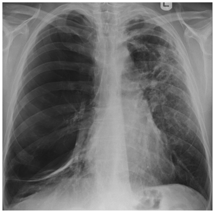
Figure 6.
Patient 2 Chest radiograph, showing extensive loss of lung architecture, 2 curvilinear opacities in the left base and the upper left mediastinum representing compressed lung from adjacent bullae formation. On the left side there is parenchymal shadowing and the impression of thin walled cavities just above the hilum.

