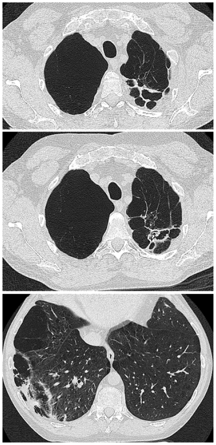
Figure 7.
Patient 2 CT scan cuts of the thorax showing extensive bullae formation on the right, cavitation posteriorly on the left, with fungal material present in a cavity on the second section, and additional areas of cavitation in the right lower lobe, with slightly thicker walls and pleural involvement.

