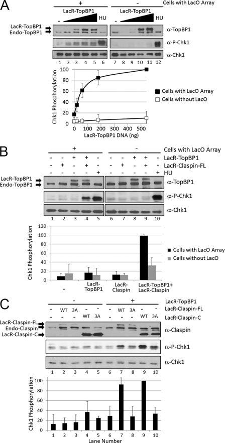
FIGURE 4.
Tethering both TopBP1 and Claspin to DNA causes synergistic induction of Chk1 phosphorylation in mammalian cells. A, LacO array-containing NIH2/4 cells (lanes 1–6) or control NIH3T3 cells (lanes 7–12) were transfected with 0, 20, 60, 180, or 540 ng of pcDNA3-LacR-TopBP1 plasmid and empty pcDNA3 plasmid such that all of the transfections had equal amounts of DNA. The cells were lysed 18 h later, and the levels of TopBP1, phospho-Chk1, and Chk1 were analyzed by immunoblotting as indicated. Quantitation of the amount of Chk1 phosphorylation after transfection is graphed below. As a positive control, cells were treated for 15 min with 2 mm hydroxyurea (HU) to induce Chk1 phosphorylation (lanes 6 and 12). B, LacO array-containing NIH2/4 cells (lanes 1–5) or NIH3T3 control cells (lanes 6–10) were transfected with 20 ng of pcDNA3-LacR-TopBP1 (lanes 3, 4, 8, and 9), 50 ng of pcDNA3-LacR-Claspin-FL (lanes 2, 4, 7, and 9), or treated with hydroxyurea (lanes 5 and 10) as in A. C, LacO array-containing NIH2/4 cells were transfected with 20 ng of pcDNA3-LacR-TopBP1 (lanes 6–10), 50 ng of pcDNA3-LacR-Claspin-FL WT (lanes 2 and 7), 50 ng of pcDNA3-LacR-Claspin-FL 3A mutant (lanes 3 and 8), 50 ng pcDNA3-LacR-Claspin-C WT (lanes 4 and 9), 50 ng pcDNA3-LacR-Claspin-C 3A mutant (lanes 5 and 10) and empty pcDNA3 as in A.

