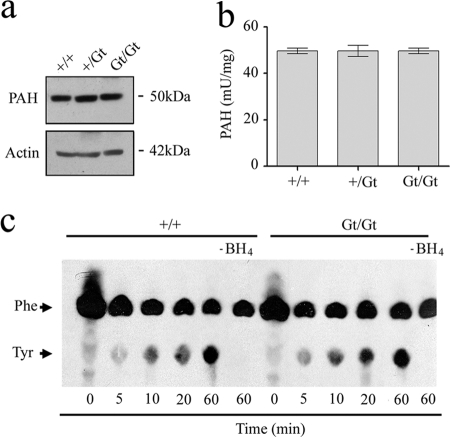
FIGURE 5.
TGT status does not influence PAH expression or activity. a, liver cytosolic extract was prepared from 6- to 8-week-old wild-type and gene-trap mice and analyzed by immunoblot using antisera against PAH and actin, the latter serving as an internal control. b, analysis of PAH activity in mouse liver extracts. Values are given per mg of total protein (n = 3 measurements from three different animals per group). c, representative figure (of three experiments) showing the measurement of [14C]phenylalanine hydroxylation in liver cytosol from wild-type and homozygous gene-trap animals that had been diluted only marginally (1:20) by the addition of phenylalanine (0.2 mm cold phenylalanine; 33 nCi [14C]phenylalanine), catalase (3.4 μg), and BH4 (100 μm). The [14C]phenylalanine and [14C]tyrosine were resolved by thin layer chromatography and visualized by autoradiography.

