FIGURE 4.
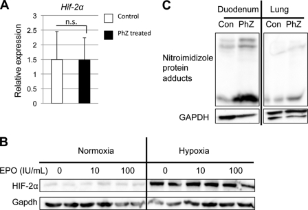
HIF-2α is stabilized by hypoxia in the small intestine following PhZ treatment. A, qPCR analysis of duodenal Hif-2α mRNA expression following PhZ or saline (Control) treatment in wild-type mice. B, Western blot analysis of Caco-2 cells treated with recombinant human EPO under normoxia and hypoxia for 24 h. Expression was normalized to GAPDH protein expression. C, Western blot analysis of nitroimidazole-protein adducts in extracts from the duodenums and lungs of PhZ-treated and control (Con) wild-type mice. Expression was normalized to GAPDH. Four to five animals for each treatment group were assessed. For Western blot analysis, a representative image from an individual mouse for each treatment group is shown. n.s., not significant.

