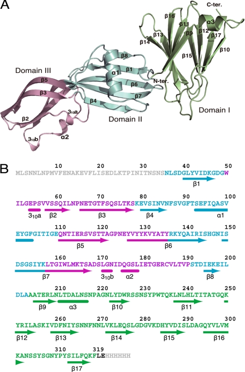
FIGURE 1.
Three-dimensional structure of the full-length CPE monomer. A, ribbon diagram of the CPE monomer. The N-terminal pore-forming domain II, domain III, and the C-terminal claudin-binding domain I are shown in blue, light pink, and lime green, respectively. B, amino acid sequence and secondary structure of CPE. Arrows and end-rounded bars below the sequence indicate β-strand and helical conformations, respectively. Domains I, II, and III are represented by green, blue, and magenta, respectively.

